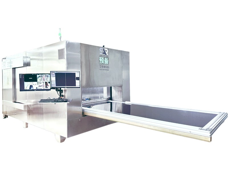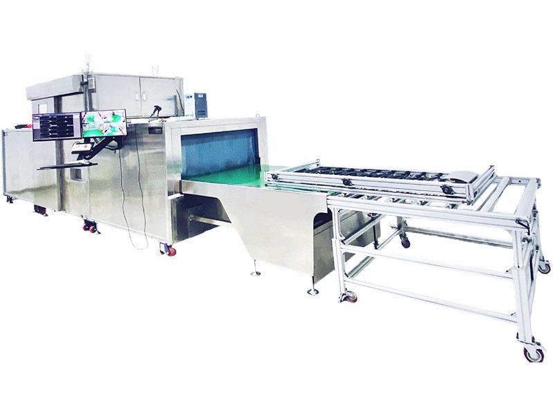


Life science and medical microscopic imaging solutions
Details
Visualization challenges in life science research
Medical Research: How to quickly and clearly image the structure of precious and vulnerable biological samples for morphological comparison, teaching display, and digital archiving?
Biological Experiments: How to obtain high-definition morphological information of the whole or specific parts of the sample without compromising its integrity, for species identification, developmental observation, or pathological screening?
High definition imaging, precise insights
The high-resolution imaging system we provide can quickly and non destructively scan various biological samples, such as small animals, plants, bones, fossils, etc. Obtaining images with high contrast and rich details can be used for morphological analysis, internal structure observation, and precise measurement, providing reliable research tools for taxonomy, comparative anatomy, developmental biology, and other studies.
Research on multi species biomorphological imaging
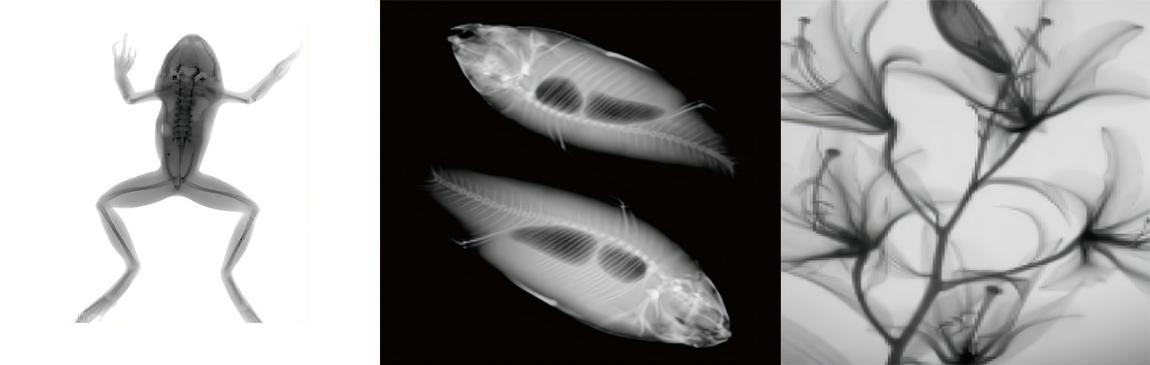
In biological research, non-destructive morphological recording of biological samples is crucial. Through our imaging system, we have successfully obtained high-definition scanning images of various biological samples. These images clearly present the internal structure and morphological features of the samples, providing accurate and intuitive imaging evidence for species classification research, teaching material production, and biodiversity research.
Explore more possibilities for sample imaging
 |
News recommendation